
- Physical complaints
- Fracture
- Stress fracture
Stress fracture
Overloading of the bone, stress fracture
Prolonged overuse of bone can lead to a fracture, a so-called stress fracture.
Bone build-up
Bones are structures in which there is normally a good balance between bone formation and bone resorption. The build-up of bone depends on the bone load, meaning that a bone becomes stronger as it is more loaded. Each load on the bone first causes minimal bone resorption and then bone formation.
Through bone formation the bone becomes stronger to cope with a higher load next time. But if the load is too high for too long and there is too little recovery, bone formation no longer occurs.
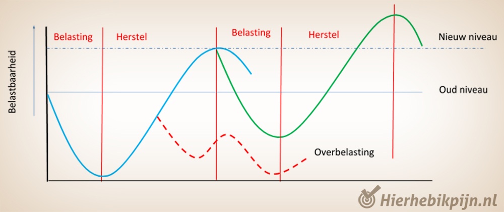
Image: After each load (bone resorption) recovery (bone formation) follows. When load is applied too soon and/or too much during recovery (red dotted line), bone formation does not proceed well. When there is insufficient recovery over a longer period, a stress fracture develops. How long this period is differs for everyone.
Risk factors for a stress fracture
There are two important risk factors for developing a stress fracture:
- Rapid increase in bone load (heavier or more training in a short time).
- Energy imbalance. With limited intake of healthy nutrients there are insufficient building blocks for bone formation.
The location of a stress fracture is sport-dependent. Stress fractures in the shoulders and arms occur in sports where the arms are used a lot, such as tennis, swimming and baseball. Athletics mainly causes stress fractures in the feet and shins. The most common sites for stress fractures are the shin, metatarsals, thigh bone and pelvis. Athletics is the sport with the greatest risk of stress fractures.
Symptoms of a stress fracture
In a stress fracture there is local pain during or after sport. The pain worsens if the athlete continues. The bone is tender to pressure at the location of the stress fracture. This applies only when the bone can be touched, such as the tibia, fibula and metatarsals. For bones that cannot be touched because there is a lot of muscle tissue around them, there are different tests to investigate whether there is a stress fracture.
Diagnosis of a stress fracture
A stress fracture has two grades:
- Low risk of worsening symptoms.
- High risk of worsening symptoms.
The location of the stress fracture determines whether it belongs to a low- or high-risk category. This depends not only on the bone, but mainly on the specific site within the bone. Thus, stress fractures in the same bone can have both low and high risk.
Imaging is necessary for a correct diagnosis. There are different options such as an X-ray, bone scan, CT scan or MRI scan.
An X-ray is not always good enough as a diagnostic tool. Stress fractures are often only visible at a later stage on an X-ray. In some cases a stress fracture is not visible at all. This is because bone changes in a stress fracture begin inside the bone. This is not visible on X-ray. An MRI scan is therefore better for diagnosing stress fractures.
Treatment of a stress fracture
In treating low-risk stress fractures, (relative) rest is sufficient. If there is sufficient pressure on the bone, such as on the thigh bone, it heals well by reducing activities. In addition, low-risk stress fractures are less likely to recur.
When a stress fracture has a high risk, the course is often unfavourable. There is a high chance that the stress fracture will become a complete fracture. Treatment in high-risk cases can be a longer period in a cast or even surgery.
Healing of the bone without surgery is often within six weeks. After this, sport can be gradually resumed. The build-up is guided by pain (stop immediately if there is pain) and tenderness when touching the fracture.
Treatment of high-risk stress fractures depends on the shape of the fracture and the location. Because of the operation, or prolonged casting, rehabilitation takes much longer. This can take up to a year.
Contact a physiotherapist to guide you in the recovery process.
What can we improve according to you?
Let us know, your opinion is important to us.
Thank you for your feedback!
We will work even harder to improve the quality of Physiocheck.co.uk.
Did this information help you?