- Conditions
- Kienböck's disease
Kienböck's disease
Lunatomalacia / avascular necrosis of the lunate bone
Introduction
Kienböck's disease is the necrosis of one of the carpal bones in the wrist. It is a small bone located between other small bones in the wrist.
Kienböck's disease is mainly seen in men between the ages of 20 and 40 and owes its name to the radiologist who first described it.
Description of the condition
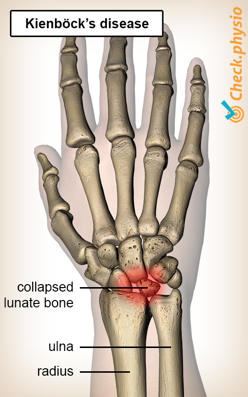
The wrist contains eight carpal bones. In the centre of these carpal bones is a moon-shaped bone called the lunate bone. Because it is located in the centre, it occupies an important place between the other carpal bones.
If there is a lack of blood supply to the lunate bone, the bone can slowly die. This process is referred to as avascular necrosis. This process causes the strength of the bone to deteriorate.
If the bone is then strained (too much), it can collapse. The other carpal bones shift as a result thereof and use the freed up space. Consequently, the hand/wrist function deteriorates and complaints of pain develop.
Due to the fact that the lunate bone necrotizes in case of Kienböck’s disease, the condition is sometimes also referred to as lunatomalacia.
Cause and origin
This condition is caused by insufficient blood supply to the carpal bone. This occurs when the blood vessels to this bone are incorrectly developed. As a result, the bone is vulnerable to damage.
Other causes include: prolonged excessive straining of the healthy bone, (micro)traumas, sickle cell disease, gout or use of cortisone.
People with Kienböck's disease regularly have a shorter ulna, the so-called ulna minus variant. This causes the forces on the bone to be greater during daily activities and the bone is therefore more heavily strained.
Phases
Four phases can be distinguished in the course of the condition. Phase 1 is already characterised by (mild) complaints, but an X-ray does not yet reveal any abnormalities. An X-ray in phase 2 shows increased density of the bone. In phase 3, the bone collapses which can lead to a change of position of the other bones. Phase 4 is clearly characterised by arthrosis (wear and tear) around the lunate bone.
Signs & symptoms
The symptoms that develop with this condition include pain and limited movement of the wrist. Depending on the stage of the condition, the following symptoms may develop:
- Pain in the wrist, especially when extending the middle finger or when exerting direct pressure on the lunate bone.
- Local swelling.
- Feeling of stiffness in the wrist joint.
- Decreased pinch strength.
- Limited movement of the wrist.
- Increased symptoms when using the hand/wrist.
Diagnosis
The doctor or physiotherapist will ask about the symptoms and what causes them. The hand and wrist are then checked in the physical examination. Tenderness of the lunate bone, combined with reduced hand function, is a clear indication that Kienböck's disease is present.
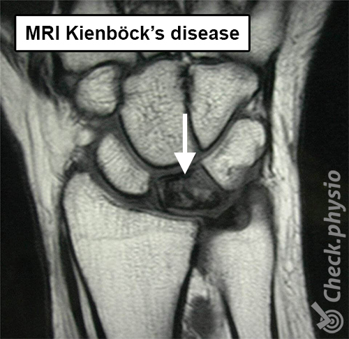
If an X-ray is taken in the early stages of the disease, it often does not yet show any abnormality. In this case, an MRI can provide more clarity. However, an X-ray at a later stage can show the condition of the lunate bone.
Treatment and recovery
When the disorder is detected relatively early, the doctor can try to reduce the complaints by immobilising the wrist using a cast or splint. If this does not help, the treatment will focus on restoring the blood supply to the lunate bone. This is achieved by transplanting (relocating) a piece of bone with accompanying blood vessel from the radius to the wrist.
If an ‘ulna minus variant’ causes the complaints, the doctor can opt to shorten the radius a bit. This way the strain on the lunate bone will decrease.
If the lunate bone is already seriously damaged, a number of options remain:
- The row of carpal bones and the lunate bone are removed entirely. This is called proximal row carpectomy.
- The wrist is secured by placing a metal plate, known as wrist arthrodesis.
- An artificial joint, called wrist prosthesis, is placed.
After surgery, a cast is worn for a few weeks. Hand) physiotherapy can start afterwards. The recovery depends on the treatment method chosen.
More info
You can check your symptoms using the online physiotherapy check or make an appointment with a physiotherapy practice in your area.
References
Nugteren, K. van & Winkel, D. (2006) Onderzoek en behandeling van de hand, het polsgewricht Houten: Bohn Stafleu van Loghum.
Verhaar, J.A.N. & Linden, A.J. van der (2005) Orthopedie Houten: Bohn Stafleu van Loghum.