- Conditions
- Lateral ankle ligament injury
Lateral ankle ligament injury Inversion injury ankle/ ankle sprain
Introduction
The lateral ankle ligament injury is a very common injury to the ankle. The symptoms usually occur after a person stumbles or sprains an ankle. If an ankle ligament injury is not treated properly, it can result in permanent instability of the ankle joint.
The injury is very common in men aged 15 - 35 years of age and in athletes. Athletes who do a lot of jumping, turning or take part in a contact sport are at increased risk.
Description of the condition
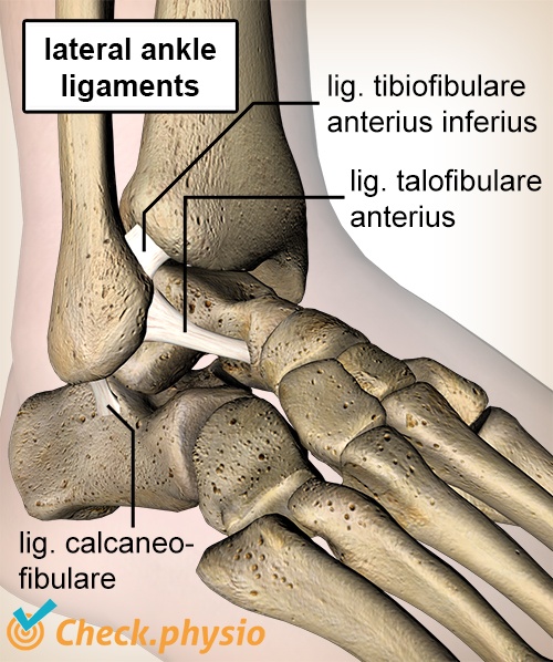
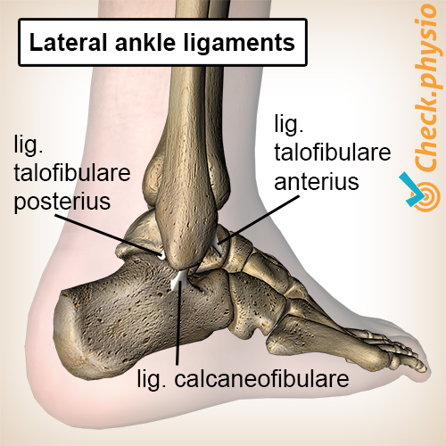
The ankle ligaments are rigid, firm band-like structures located on the inside and the outside of the ankle. They connect the lower leg to the foot and absorb the shock of extreme movements in the ankle joint.
When we stumble or sprain an ankle, this causes maximum stretching of the ankle. In most cases they prevent the ankle from folding over completely. If the forces are greater than the ankle ligaments can take, they can tear completely or partially.
The injury is categorised into various grades. A 'grade 1' injury is a mild sprain. Only a few fibres in the ligament are torn. A 'grade 2' injury is more serious and one of the ligaments can tear completely (total rupture). If several ligaments are torn, resulting in the ankle joint becoming completely unstable, this is referred to as a 'grade 3' ankle ligament injury.
Cause and origin
The symptoms usually occur at a clearly defined moment when a person stumbles or sprains the ankle. In some cases, the ankle ligaments can also become overloaded through prolonged stretching during sports or other activities.
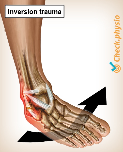
The typical sprain that causes injury to the lateral ankle ligaments is also referred to as the 'inversion trauma'. In this injury, the sole of the foot rotates inwards and the ankle rotates outwards (refer to image). The lateral ankle ligaments are fully stretched as a result.
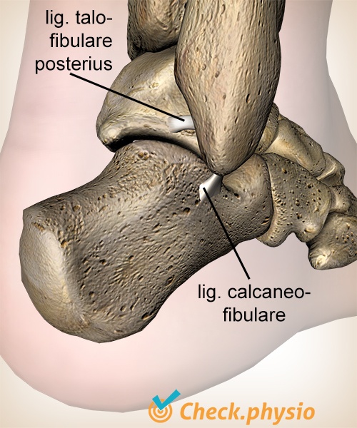
In general the ankle ligaments tear in the same order. For example, the ankle ligament called the 'anterior talofibular ligament' is usually the first ligament affected. The following ligaments to tear - in order - are the 'calcaneofibular ligament', the 'posterior talofibular ligament' and the 'anterior inferior tibiofibular ligament'.
Signs & symptoms
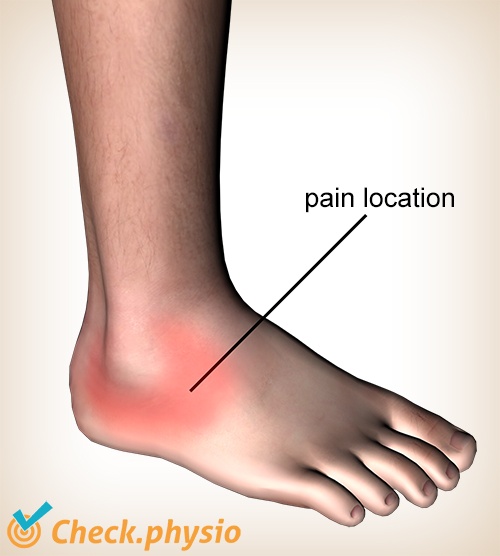
- Pain on the outside of the ankle/foot.
- Swelling (immediately and in the first few days after the injury).
- Sometimes a bruise (haematoma) can be present.
- Feeling of instability in the ankle.
- Almost all movements of the ankle can be painful. In particular, the (passive) inward rotation of the base of the foot (inversion) makes the symptoms worse, as this stretches the lateral ankle ligaments.
Diagnosis
The diagnosis is confirmed by means of the physical examination. An ankle fracture should be considered if the patient is unable to walk more than four steps on the affected ankle.
Treatment and recovery
The treatment depends on the severity of the injury. The physiotherapist can examine this. Often it is sensible to walk on crutches for the first few days after the injury, to reduce strain on the torn ligaments. An pressure bandage is prescribed and the leg should be elevated. Once the swelling has partly gone down, the ankle can be taped.
Treatment consists of exercises for the ankle that aim to improve the mobility, strength and stability. The strain is gradually increased to the level at which the patient wants to be able to function (activities or sport). If the patient repeatedly sprains the ankle, one can consider using an ankle brace.
One should take into consideration that complete recovery of sprained or torn ankle ligaments takes a minimum of 6 weeks and may even take longer than a year. Allowing the injury to heal without intervention can result in chronic instability. This increases the risk of repeated sprains by three-fold.
Exercises
During the recovery process, it is important to stimulate the ankle ligaments in a responsible manner so that they become strong again. View a professionally compiled exercise programme here with exercises for the lateral ankle ligament.
More info
You can check your symptoms using the online physiotherapy check or make an appointment with a physiotherapy practice in your area.
References
Nugteren, K. van & Winkel, D. (2009) Onderzoek en behandeling van de voet Houten: Bohn Stafleu van Loghum.
Verhaar, J.A.N. & Linden, A.J. van der (2005) Orthopedie Houten: Bohn Stafleu van Loghum.