- Conditions
- Pes anserinus syndrome
Pes anserinus syndrome Pes anserinus tendinopathy, pes anserinus bursitis
Introduction
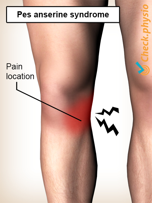
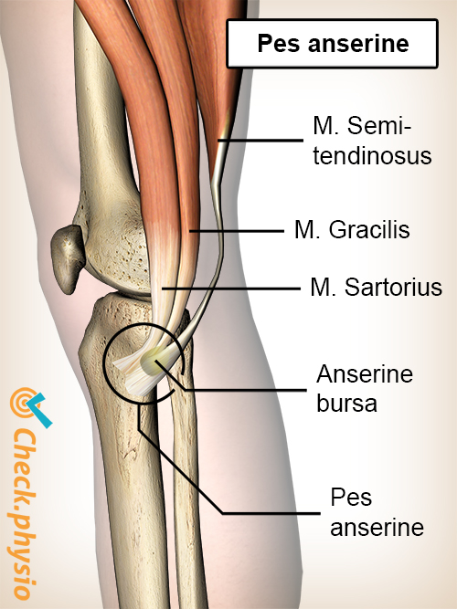
The pes anserinus is the insertion of three tendons on the medial side of the knee joint. Literally, pes anserinus means "goose foot", due to the shape of these three tendons. Pain in the pes anserinus can be caused by inflammation of one of the tendons or by bursitis. The pain is felt on the medial side of the knee.
Description of the condition
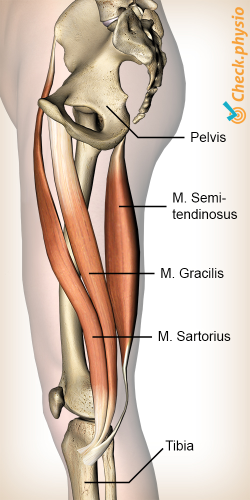
The pes anserinus is formed by the tendons of three separate muscles: the gracilis, sartorius and semitendinosus. These tendons attach to the tibia. The function of these three muscles is mainly to bend and rotate the knee and to provide support in order to prevent valgus stress. Valgus stress is the inward caving of the knee, which is why it is also referred to as a knock knee. The space between this conjoined tendon and the tibia contains a bursa. A bursa is a bag filled with fluid that makes sure that different structures slide over one another.
The difference between tendinitis (tendinopathy) or bursitis is difficult to be distinguish. Hence, the term pes anserinus syndrome is used. The difference also has no added value because the treatment is the same in both cases.
Cause and origin
An irritated, damaged or infected bursa or tendon causes complaints. In this case, the bursa causes that the tendons cannot slide well with respect to the tibia.
Primarily the position of the knee (knock knee) causes complaints in the pes anserinus. Because of the changed position, the tibia can exert more pressure on the bursa and tendons of the pes anserinus.
Additionally, tight hamstrings also cause complaints in the pes anserinus. This is because one of the tendons, (the semitendinosus), is a tendon of one of the hamstrings.
In turn, tight hamstrings and the knock knee can have different causes. Besides the built of the body, wear and tear of the knee joint and meniscus problems constitute a risk of developing knock knee. Increased tension on the hamstrings can be caused by incorrect posture. The hamstrings are attached to the pelvis so that the position of the pelvis is decisive for the mobility of the hamstrings.
Risk factors for developing pes anserinus complaints
- Female gender (the pelvis is usually wider, developing into knock knee faster).
- Obesity (the pressure on the knees becomes too high and this can cause knock knee).
- Arthrosis of the knee (which can cause knock knee).
- Diabetes (reduced circulation so that tendons and bursas recover less quickly).
- Rheumatism (reduced quality of the muscles results in higher likelihood of wear and tear).
- Repetitive sports (running, cycling, swimming can cause excessive strain).
- Flatfeet (which can play a role in the position of the knee).
- Local trauma (accident, fall or making a wrong movement).
- Osteophyte formation (extra ossification on the medial side of the knee).
Signs & symptoms
Typical symptoms include pain on the medial side of the knee and pain when pressing on the insertion of the tendons. The insertion sits below the knee on the medial side of the tibia. The pain often gets worse by walking up and down the stairs. The typical symptoms are not always present, and the location of the pain can be different for everyone. Pain is often experienced over a larger area. As a result, it is difficult to distinguish if the pain originates from the pes anserinus, medial meniscus or medial knee ligament.
The set criteria for the diagnosis include:
- Pain on the medial side of the knee.
- Pain when walking up and down the stairs.
- Pain in the morning.
- Morning stiffness at the location (of the pain) lasting at least one hour.
- Difficulty getting out of a chair or car.
- Local sensitivity.
- Local swelling.
Diagnosis
The diagnosis is made by a (general) practitioner or physiotherapist on the basis of an interview and physical examination. The above-mentioned criteria play an important role in this. Additional examinations such as X-ray examination or ultrasound examination have no added value in the case of these symptoms.
An X-ray examination can, however, be used to determine the degree of osteoarthritis in the knee. Osteoarthritis is a risk factor for the development of pes anserinus symptoms.
An ultrasound examination identifies muscles, tendons and bursas, but it is known that this is not a reliable examination when it comes to pes anserinus symptoms.
When making the diagnosis, other problems in this region must be ruled out. Mainly meniscus problems and medial ligament damage cause pain in the same region. This should be taken into account when making the diagnosis.
Treatment and recovery
The treatment of pes anserinus syndrome focuses on reducing the inflammatory reaction. This can be achieved by means of (relative) rest, icing and anti-inflammatory drugs prescribed by the (general) practitioner. A local injection of anti-inflammatories may be administered. To sleep, the use of a pillow between the knees is advised to reduce pressure on the painful spot.
In addition, the treatment consists of tackling the risk factors. If overweight, the patient is urgently advised to lose weight. If the patient has flatfeet, these can be treated with insoles.
Physiotherapy plays an important role during treatment. The physiotherapist can treat the muscles around the knee joint through exercises and certain stretching techniques. Posture is worked on too, especially when the patient has knock knee. If necessary, strength training is advised and given to prevent loss of muscle strength around the knee joint.
More info
You can check your symptoms using the online physiotherapy check or make an appointment with a physiotherapy practice in your area.
References
Helfenstein, M. & Kuromoto, J. (2010) Anserine syndrome Bras / Rheumatol. 2010;50(3):313-27.
Mohseni, M. & Graham, C. (2018) Bursitis, Pes Anserine StatPearls Publishing; 2019 Jan.