- Conditions
- Pelvic instability
Pelvic instability Pelvic pain / pelvic girdle pain / PGP
Introduction
The pelvic ring is very important in the transfer of forces between the trunk and the legs. The ring is formed by various bone segments and joints. Any instability in the joints can cause problems, which is called pelvic instability.
Pelvic instability is common in pregnant women and women who have recently given birth. Nevertheless, men and non-pregnant women can also be affected.
Description of the condition
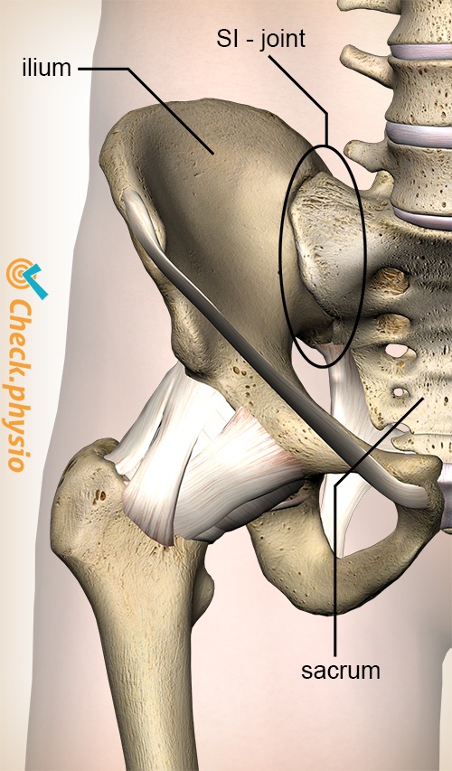
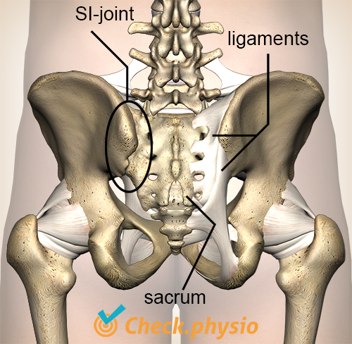
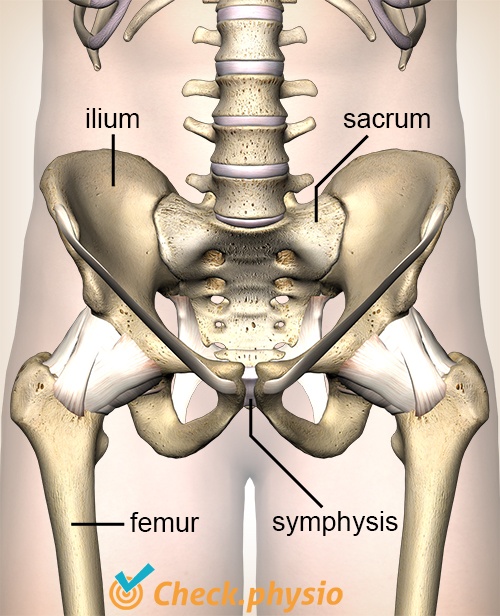
The pelvis comprises three bone segments: the left pelvic half, the right pelvic half and the sacrum. The sacrum is wedged between the two pelvic halves and is located at the rear of the body. The pelvic halves are connected to each other along the front of the body. These bone segments together form a circular shape called the pelvic ring.
The bone segments are connected to each other by means of three joints. At the rear these are the sacro-iliac joints to the left and right of the sacrum (abbreviated: SI joints). At the front there is a third joint. This is called the 'pubic symphysis'. Various ligaments running over the joints keep these bone segments together.
When the ligaments slacken the joints become more mobile. This can be very functional, as is the case in pregnancy. However, this can also cause problems as a result of overloading or damage to the ligaments. This is referred to as pelvic instability.
Cause and origin
The symptoms usually occur during pregnancy or shortly after giving birth. The hormone 'relaxin' causes the ligaments to soften and the pelvic joints to become more mobile. This allows the pelvis to change shape more easily and makes it easier for the child to pass through the pelvic ring during birth.
It is not only (pregnant) women who are affected by pelvic instability. A slackening of the transverse and oblique abdominal muscles can also cause these symptoms. In addition, an accident or fall whilst playing sport, whilst working or in traffic can cause over-stretching or tearing of the pelvic ligaments, resulting in pelvic instability.
Signs & symptoms
The presentation can differ per patient. The symptoms listed below are observed frequently:
- Pain in the lower back, the coccyx, the abdomen, along the side or rear of the thigh, in groin or the pubic bone.
- Problems with activities such as walking on stairs, rolling over in bed, prolonged sitting, getting up from a chair or walking.
- Pain with jolting movements (for example, when running or cycling over a bumpy road).
- Pain during menstruation or intercourse.
- Inability to stand on 1 leg.
- The pain can increase after a day with a lot of activities.
- Start-up pain: more pain when moving after a prolonged period of inactivity (for example when getting up in the morning).
- Fatigue.
Treatment and recovery
Appropriate information is very important. The midwife, gynaecologist and (pelvic floor) physiotherapist play an important role here. The treatment can consist of the use of a pelvic band, physiotherapy, exercises to perform at home, fitness training and training of the muscles.
The patient must also learn to cope with the symptoms. A good balance between rest and activity is very important. The body needs rest to allow the capsules and ligaments to recover, but also needs movement to prevent muscle weakness. Acceptance also plays an important role in this. There is no 'miracle cure' as yet for pelvic instability.
Exercises
Follow the exercise programme with exercises for pelvic instability. The specially composed exercises in this programme are aimed at training the stability of the pelvis.
More info
You can check your symptoms using the online physiotherapy check or make an appointment with a physiotherapy practice in your area.
References
El, A. van der (2007) Manuele diagnostiek. Wervelkolom Rotterdam: Uitgeverij Manthel.
Kleiverda, G. (2001) Bekkenpijn, bekkeninstabiliteit en zwangerschap Voorlichtingsbrochure. Nederlandse Vereniging voor Obstetrie en Gynaecologie.