- Pain in the knee.
Pain in the knee.
Various structures in and around the knee work together to keep the knee both stable and mobile. Almost everyone experiences knee problems at some point. Knee complaints can arise suddenly or develop gradually over a longer period. This often manifests as pain, stiffness, restricted movement, or a swollen knee.
Anatomy of the knee
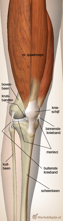
The structure of the knee can be confusing for many people. Terms such as 'meniscus', 'knee ligaments', and 'cruciate ligaments' are often used interchangeably. Below is an overview of the main structures in and around the knee.
Thigh bone and shin bone
Different bones meet in the knee joint. The rounded joint surfaces of the thigh bone (the femur) rest on the flatter top of the shin bone (the tibia). Together they form the 'tibiofemoral joint'. Because the joint surfaces of the thigh bone do not fit perfectly on the flat 'tibial plateau', there are two menisci (plural of 'meniscus') between the bone ends.
Meniscus
A meniscus is a crescent-shaped cartilage disc that helps distribute pressure in the knee joint and acts as a shock absorber. It is also thought that the menisci contribute to better lubrication of the joint by optimally guiding the joint fluid (synovia) along the joint cartilage.
Cartilage
The joint cartilage is an extremely smooth layer of connective tissue that covers the bone surfaces that glide over each other. Wear of this cartilage layer is also known as 'arthrosis' (joint wear).
Ligaments
The stability of the knee is largely provided by various ligament structures. They hold the knee firmly together without restricting its natural movements.
Between the inner (medial) and outer (lateral) meniscus are the anterior and posterior cruciate ligaments. These should not be confused with the inner (medial) and outer (lateral) collateral ligaments, which are located on either side of the joint.
Kneecap
The 'patella', better known as the kneecap, protects the front of the knee and plays an important role in helping the quadriceps muscle transfer forces to the lower leg. The patella forms a separate joint with the femur, also known as the 'patellofemoral joint'.
Fibula
On the outside of the knee is a small joint formed by the head of the fibula firmly connected to the tibia. This is called the tibiofibular joint (tibia = shin bone, fibula = calf bone).
Muscles
The muscle that runs from the hip along the front of the thigh and then via a tendon attaches with the kneecap to the lower leg is the quadriceps muscle. This muscle functions to extend the knee.
At the backside of the knee and the thigh are the hamstrings. The main function of the hamstrings is to bend the knee. Together with many other muscles, the quadriceps and hamstrings work closely to allow smooth rotation, bending, and extension of the knee.
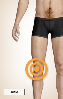
Check your knee complaints
To get a good impression of what may be causing the pain in your knee, we recommend that you do the online physiotherapy check. By indicating where you feel pain and then answering the questions, your knee complaints are compared with the most common conditions within physiotherapy. It’s wise to have your knee pain checked by a physiotherapy practice in your area.
Knee disorders
Below is an overview of conditions that can cause pain in the knee. Each article provides detailed information about the complaints, the cause, and possible treatment. When you do the online physiotherapy check, your complaints are compared with all these conditions.
Knie
- Acute hamstring injury
- Acute meniscus injury
- Anterior cruciate ligament injury
- Baker's cyst
- Bruising
- Degenerative meniscus injury
- Fibromyalgia
- Hamstring injury
- Hoffitis
- Iliotibial band syndrome
- Joint mouse
- Jumper's knee
- Lateral knee ligament injury
- Medial knee ligament injury
- Myositis ossificans
- Osgood-Schlatter
- Osteoarthrosis of the knee
- Osteochondritis dissecans
- Patellofemoral pain syndrome
- Pes anserinus syndrome
- Popliteus tendinitis
- Posterior cruciate ligament injury
- Prepatellar bursitis
- Quadriceps tendinitis
- Rheumatoid arthritis