- Anatomy
- Anatomy of the wrist
Anatomy of the wrist
The wrist spans from the forearm to just before the fingers. It comprises bones, capsules, ligaments, muscles, tendons, nerves, and blood vessels, all working together to enable smooth hand movement and weight-bearing support.
Bones
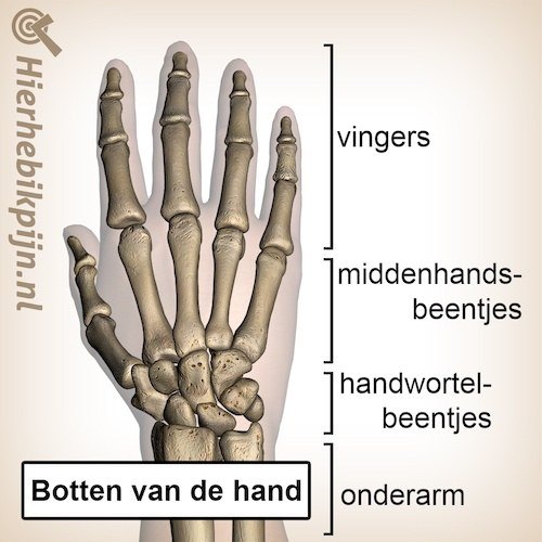
The forearm consists of two bones, the radius and the ulna. The ends of these two bones form one half of the wrist, while the other half is formed by the carpal and metacarpal bones, which are part of the hand.
Between these bones lies the triangular fibrocartilage complex (TFCC), which includes a cartilage disc, ligaments, and a tendon sheath. Together, these structures fill the space between the ulna and the carpal bones. Additionally, they connect the ulna, radius, and one of the carpal bones (os triquetrum), providing the wrist with stability and helping to absorb forces.
Capsule and ligaments
A significant portion of the wrist's stability comes from the surrounding capsule and ligaments, which are divided into two groups: intrinsic and extrinsic ligaments.
Intrinsic ligaments are short and stiff, connecting one carpal bone to another. Extrinsic ligaments are longer, originating from the forearm bones and extending across multiple bones. These ligaments are slightly more flexible, allowing for some movement. Together, they form a network of ligaments and bands that provide both stability and mobility to the wrist.
Muscles
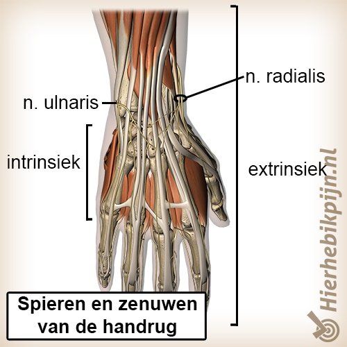
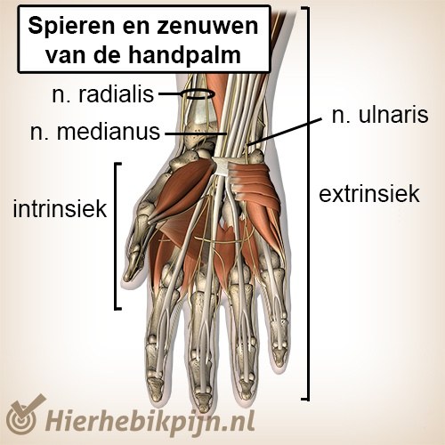
The muscles responsible for wrist movement are primarily located in the forearm. From there, they extend via long tendons across the wrist. The muscles that flex the wrist are located on the palm side, while those that extend the wrist are situated on the back of the hand.
Nerves
The nerves that control muscle movement and provide sensation in the hand also pass through the wrist. The three main nerves found here are the ulnar, radial, and median nerves.
The median nerve, along with nine tendons, runs through the carpal tunnel, a small tunnel on the palm side of the wrist formed by the carpal bones and a ligament. This area plays a crucial role in carpal tunnel syndrome.
Blood vessels
Two important blood vessels, the ulnar and radial arteries, run across the wrist to the fingers. The radial artery is commonly used to measure the pulse.
Related topics
- Subscapularis
- Connective tissue
- Joint
- Ligaments
- Joint capsule
- Synovial fluid
- Bursa
- Nervous system
- Central nervous system
- Peripheral nervous system
- Motor nerve
- Sensory nerve
- Muscles
- Tendons
- Proprioception
- Dermatomes
- Hamstrings
- Achilles tendon
- Anatomy of the wrist
- Anatomy of the hand
- Osteoarthritis
- Anatomy of the foot
- Big toe
- Trigger points
- Anatomy of the thigh
- Cartilage
- Rotator cuff
- Intervertebral disc
- SI joint
- Lungs
- Anatomy of the abdominal muscles
- Anatomy of the bones
- Pelvic floor muscles
- Trapezius muscle
- Skin
- Anatomy of the spine