- Anatomy
- Anatomy of the foot
Anatomy of the foot
The foot is a complex body part composed of various structures that must work together seamlessly and move correctly to enable activities such as walking and jumping. It also plays a crucial role in maintaining stability during standing and movement, requiring a balance between stability and mobility.
Important structures in the foot are: bones, muscles, joint capsules, ligaments, nerves and blood vessels. The foot has thirty-three joints in total that ensure mobility. Around each joint there is a joint capsule with ligaments.
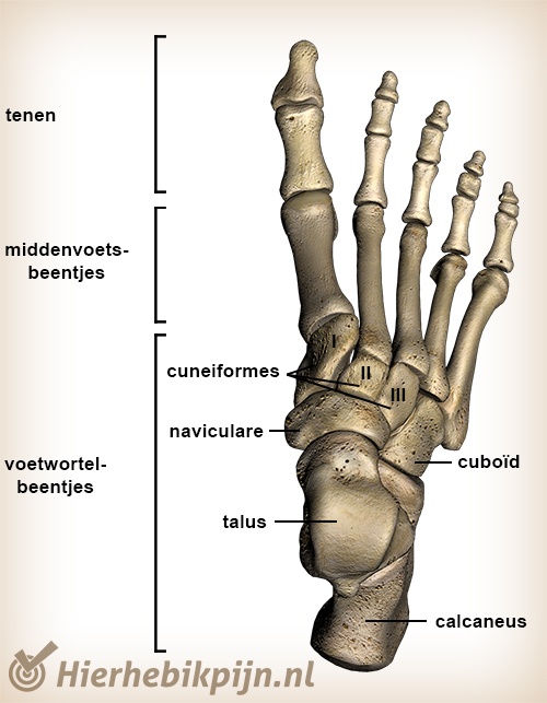
Bones of the foot
The foot consists of:
- Toes (phalanges).
- Midfoot (metatarsus).
- Hindfoot (tarsus).
Toes
The toes are made up of fourteen small bone segments forming five toes. These bones are anatomically similar to finger bones but much shorter. Each toe consists of three bones, except for the big toe, which has two.
Midfoot
The midfoot contains five metatarsal bones (metatarsalia), labeled with Roman numerals I to V, where I is the big toe and V is the little toe.
Hindfoot
The hindfoot consists of seven bones:
- Os talus.
- Os calcaneus.
- Os naviculare.
- Os cuboideum.
- Os cuneiforme mediale.
- Os cuneiforme intermedium.
- Os cuneiforme lateralis.
The two largest bones, the talus and the calcaneus, bear most of the body's weight and form the pivot point of the ankle. Together, they create the subtalar joint.
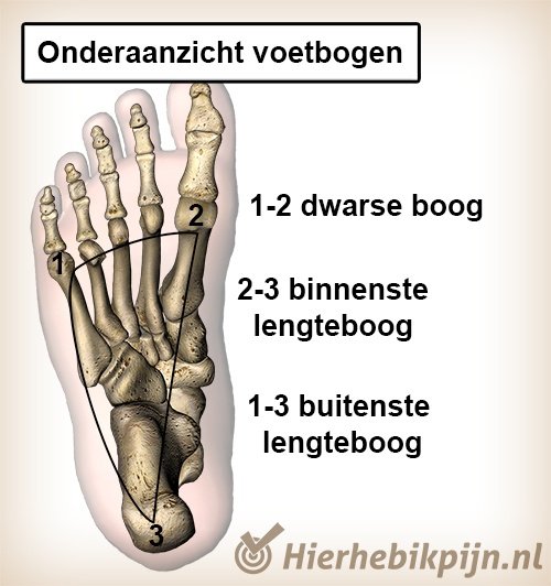
Foot arches
The foot features three foot arches, shaped by the bones, ligaments, and muscles. When ligaments are taut, the foot acts as a stiff unit; when relaxed, it moves more flexibly to absorb shocks.
Two arches run longitudinally along the foot: one on the inner side and one on the outer side, forming the foot's hollow. The third arch runs transversely across the metatarsal bones just before the toes.
Function of the foot arches
The arches consist of various foot bones forming different joints. These joints work together to create a coupled movement system in the foot. If one joint has issues, another can compensate by adjusting its movement.
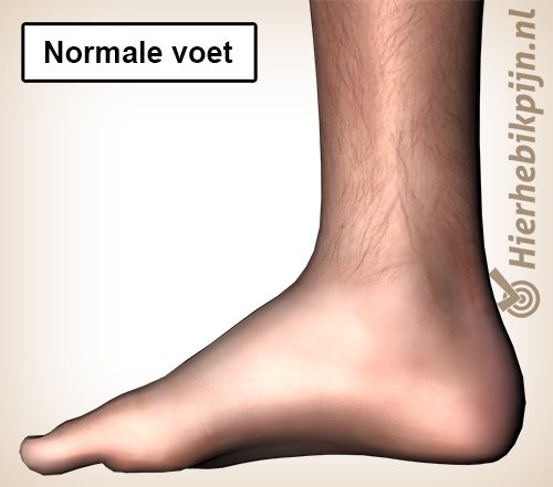
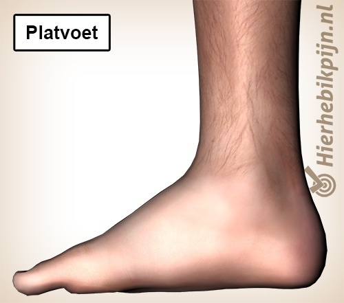
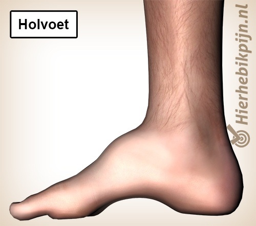
The shape of the foot arches is determined by the foot's structure, while ligaments and muscles control their function. Many arch abnormalities, such as flat feet (pes planus) and high-arched feet (pes cavus), can be compensated for, except those caused by structural problems.
Movements of the foot
The foot primarily moves from the ankle joint, with the only active movements in the foot itself being the flexion and extension of the toes.
Many movements occur between the metatarsal and hindfoot bones, which are crucial for absorbing shocks, such as walking on uneven surfaces. For example, walking on the beach causes movement between the metatarsal bones to maintain optimal foot arch shape. It is impossible to move metatarsal III separately from metatarsal IV consciously.
The foot can move in three directions via the ankle, usually in combination:
Foot flexion and extension
- Plantarflexion (standing on the toes).
- Dorsiflexion (drawing the toes toward you).
Sideways movement of the foot
- Abduction (moving the foot outward).
- Adduction (moving the foot inward).
Tilting of the foot
- Supination (tilting the foot inward).
- Pronation (tilting the foot outward).
A well-known combination of movements that often leads to injury is an ankle sprain (inversion trauma), involving simultaneous plantarflexion, adduction, and supination.
Foot complaints
Foot complaints can result from incorrect movements or overloading structures. For an overview of foot-related issues, visit the page on pain in the foot. If you need help addressing foot complaints, consider making an appointment with a physiotherapist near you.
Related topics
- Subscapularis
- Connective tissue
- Joint
- Ligaments
- Joint capsule
- Synovial fluid
- Bursa
- Nervous system
- Central nervous system
- Peripheral nervous system
- Motor nerve
- Sensory nerve
- Muscles
- Tendons
- Proprioception
- Dermatomes
- Hamstrings
- Achilles tendon
- Anatomy of the wrist
- Anatomy of the hand
- Osteoarthritis
- Anatomy of the foot
- Big toe
- Trigger points
- Anatomy of the thigh
- Cartilage
- Rotator cuff
- Intervertebral disc
- SI joint
- Lungs
- Anatomy of the abdominal muscles
- Anatomy of the bones
- Pelvic floor muscles
- Trapezius muscle
- Skin
- Anatomy of the spine